News
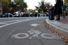
Cambridge Residents Slam Council Proposal to Delay Bike Lane Construction

News
‘Gender-Affirming Slay Fest’: Harvard College QSA Hosts Annual Queer Prom

News
‘Not Being Nerds’: Harvard Students Dance to Tinashe at Yardfest
News
Wrongful Death Trial Against CAMHS Employee Over 2015 Student Suicide To Begin Tuesday

News
Cornel West, Harvard Affiliates Call for University to Divest from ‘Israeli Apartheid’ at Rally
X-Rays Travel From Newton To Boston by Computer
Radiologists Use Teleradiography to Transmit Images Between Offices, Hospitals and Homes
In an emergency, Dr. Gerald M. Kolodny, director of nuclear medicine at Beth Israel Hospital in Boston, doesn't need to leave his Newton home to examine his patients' X-rays.
And Kolodny, an associate professor of radiology at the Medical School, is just one of many area radiologists who are excited about the latest fusion of state-of-the-art computer technology and medicine, a field known as teleradiography.
Teleradiography allows physicians to use computer networks to to share images with colleagues and students around the area, thus making possible efficient collaborations on diagnoses and treatment programs. By digitizing data from magnetic resonance imaging (MRI), CAT scans and some digital X-ray machines, radiologists such as Kolodny can transmit X-rays of broken bones all over the world.
The use of teleradiography requires just a standard UNIX terminal connected to the internet.
Dr. Justin D. Pearlman '75, director of magnetic resonance imaging (MRI) technologies and computing at Beth Israel and founder of Unified Medical Systems, a Brookline company which creates and utilizes digital images, is now heading up the effort at Beth Israel to make the use of teleradiography more widespread.
Presently, there is a limited use of teleradiography in the hospital's radionuclear department. But Pearlman, who is also a member of the Harvard-MIT Division of Health Sciences and Technology, says he hopes more doctors will soon use it.
"We use it nearly every day and send medical images all over the world," says Pearlman, who, like Kolodny, receives images at his home in the case of emergencies.
Other area hospitals, including Harvard affiliates, are also beginning to make use of the new technology. At Brigham and Women's Hospital, radiologists are currently experimenting with network-linked MRIs and CAT scans.
According to Ethan F. Fener, director of radiology information systems at Brigham and Women's, of nearly 200,000 radiology exams performed each year, 30 percent are digital.
"Nearly all of those exams are written onto various computer file servers and are available for transmission through the department, the hospital, and in some instances, the medical area," he said.
For example, CAT scans are performed at Brigham and Women's and transferred via the network to the nearby Dana Farber Cancer Institute, where radiation therapy is planned and sent back to the hospital to be executed.
But at some hospitals, physicians are shying away from the technology, citing disadvantages including expense and speed.
At Massachusetts General Hospital (MGH), doctors primarily focus on picture archiving, which allows digital images to be stored in large quantities, instead of teleradiology, according to Pearlman.
And a radiology department employee at Children's Hospital, who would not give her name, said that the hospital does not use teleradiography.
"At this point, we are acquiring images without destructive compression, but speed becomes an issue," said Michael A. Viera, assistant director of radiology administration at Brigham and Women's.
According to Pearlman, additional disadvantages involve the inconveniences of computer terminals. For example, he says, some computer screens are limited both in the amount of space available at a time, the intensity and the lack of brightness available.
But a recent study by one MGH radiologist found that images transmitted between the hospital and an outpatient community health center in Burlington, Mass. were interpreted the same in 97 percent of cases by radiologists.
Fener says the only disadvantage is the expense involved in expanding the system, and that demand for the service "is overwhelming."
And Pearlman says that the advantages to using the teleradiography system justify the cost.
"We can extract things that may not be all that apparent to the eye," says Pearlman. "It allows you to integrate information from different images and view things in three-dimensions, and you can look at the texture and intensity ranges [that are otherwise impossible to see]."
According to Fener, future applications of teleradiography will include making images more widely accessible to physicians and using the data for analysis, therapy and intervention.
"Physicians will learn whatever they need to learn," says Pearlman. "It needs to be the case that they'll get the best answer. We've just introduced new tools that will make that the case."
Want to keep up with breaking news? Subscribe to our email newsletter.